The PDF file of the papar on this topic
(IEEE Transactions on Visualization and Computer Graphics, 6(2): 160-180 2000)
is available
(click here for PDF file!).
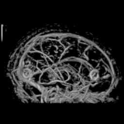
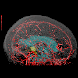
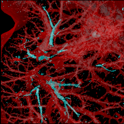
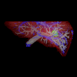
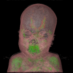
All the images in this page were created using
the volpack library developped by P. Lacroute and M. Levoy at Stanford University.
Yoshinobu Sato is with
the Division of Interdisciplinary Image Analysis,
Osaka University Medical School.
The volume rendered images were partly created while he
was staying as a visiting researcher
at
the Surgical Planning Laboratory (Director: Ron Kikinis),
Department of Radiology,
Brigham and Women's Hospital and Harvard Medical School in 1996 and 1997.
Collaborators:
Carl-Fredrik Westin,
Abhir Bhalerao,
Shin Nakajima,
Nobuyuki Shiraga,
Shigeyuki Yoshida,
Gary Zientara, and
Ron Kikinis.
Acknowledgement: Yoshi Sato would like to specially thank Shin Nakajima
because his volume rendering images created by Macintosh inspired me
to do this work and he has been continuously giving me the inspiration of the research.
Return to Yoshi's Home Page