Yoshinobu Sato's Volume Rendering Gallery
of 3D Medical Images
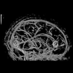
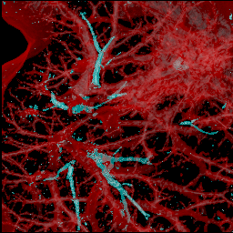
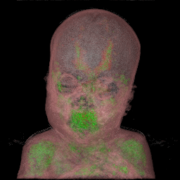
Tissue Classification Based on 3D Local Intensity Structures
for Volume Rendering
Abstract:
3D image filters for
the enhancement of specific local intensity structures such as line and sheet,
and their application to tissue classification for volume rendering are demonstrated.
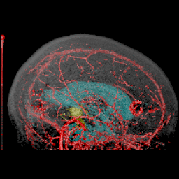
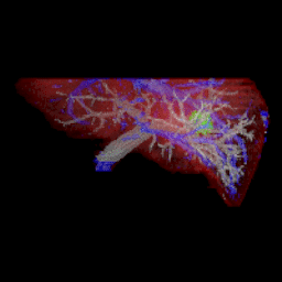
Multi-channel classification is performed
by combining different 3D image filter outputs.
The resulted method
significantly enlarges the scope of volume rendering, especially in the medical domain.
We show the usefulness of the method for various visualization
problems.
Requirements for Visualization of 3D Medical Images
Minimize Burden of Interactive Segmentation
Maximize Objectivity and Reality
Our Approach: 3D Image Filter + Volume Rendering
3D Image Filters for Enhancement of Specific Tissues
Multi-Channel Tissue Classification by Combining Different 3D Image Filter Outputs
The postscript file of the papar on this topic
(in Proc. JAMIT Frontier '97, Suita, Japan, pp.167-172, Jan 1997)
is available
(click here for PS file!).
All the images were created using
the volpack library developped by P.Lacroute and M.Levoy at Stanford University.
Collaborators:
Carl-Fredrik Westin,
Abhir Bhalerao,
Shin Nakajima,
Nobuyuki Shiraga,
Shigeyuki Yoshida,
Gary Zientara, and
Ron Kikinis.
Acknowledgement: Yoshi Sato would like to specially thank Shin Nakajima
because his volume rendering images created by Macintosh inspired me
to do this work and he has been continuously giving me the inspiration of the research.